What Is a Corneal Dystrophy?
Corneal dystrophies are inherited diseases of the cornea that often progress or worsen over time. They cause decreased vision by causing the normally clear, smooth cornea to become cloudy or irregular. There are many different types of dystrophies and they are usually categorized according to the layer of the cornea they affect. The cornea can be divided into three important layers:
- Front Layer (Epithelium)
- Middle Layer (Stroma)
- Back Layer (Endothelium)
 Front Layer
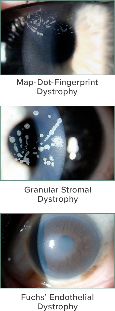
Dystrophies that affect the front layer or epithelium can cause the corneal surface to be irregular. A normal cornea has a smooth surface and any irregularities can cause blurriness or fluctuating vision. The most common front layer or epithelial dystrophy is called basement membrane dystrophy or Map-dot-fingerprint dystrophy.
Middle Layer
Dystrophies that affect the middle layers or stroma are rare and usually inherited. They usually make the normally clear cornea cloudy. The cloudy opacities in the cornea reduce vision. The most common stromal dystrophies are called lattice, granular and macular dystrophy.
Back Layer
Dystrophies that affect the back or innermost layer of the cornea called the endothelium usually cause the cornea to swell. The function of the endothelial layer is to pump fluid out of the cornea. If it is damaged, the cornea swells and the vision becomes cloudy. The most common dystrophy to affect the back layer is Fuchs’ dystrophy.
Map-Dot-Fingerprint Dystrophy
How Is a Corneal Dystrophy Diagnosed?
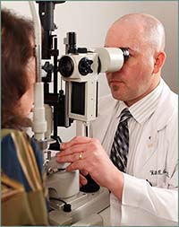 Most corneal dystrophies are easy to diagnose during a routine eye exam using a special microscope designed to look at the eye called a slit lamp. Corneal dystrophies look like cloudy opacities in the normally clear cornea or irregularities on the corneal surface. In some cases a corneal topography may aid in the diagnosis. A corneal topography is like a topographical map of the cornea that shows if the surface is misshapen or irregular.
How Are Corneal Dystrophies Treated?
The cornea trained specialists at Northeast Ohio Eye Surgeons can diagnoses a corneal dystrophy and have many treatments available to help improve and restore vision. If a dystrophy is mild, it may not cause any symptoms. The vision in patients with milder dystrophies can sometimes be corrected with glasses or contact lenses. Sometimes specialty contact lenses like RGP (Rigid Gas Permeable) or scleral lenses may be useful. These lenses help to smooth the surface of an irregular cornea.
If a dystrophy is severe, a surgical treatment may be recommended. The surgical treatment for a dystrophy often depends on which layer of the corneal is affected.
Front Layer
Dystrophies that affect the front layer or epithelium can be treated by removing the affected layer with a procedure called a superficial keratectomy. The epithelial layer has the remarkable capacity to regenerate and the new layer that replaces the old one is often much clearer and smoother.
Middle Layer
Dystrophies that affect the middle layers of the cornea are more difficult to treat. If the corneal cloudiness is closer to the surface a PTK (phototherapeutic keratectomy) may be performed. This is laser procedure that can remove opacities and smooth the cornea. If the cloudiness is deep in the cornea the best treatment may be a cornea transplant, such as DALK and PK. DALK (Deep Anterior Lamellar Keratoplasty) preserves the delicate innermost layer of the cornea, and provides a speedier recovery and prevents transplant rejection.
Back Layer
Dystrophies that affect the back or innermost layer of the cornea called the endothelium may need no treatment there is minimal or no swelling in the cornea. Advanced forms of corneal transplant such as DSAEK or DMEK are the best options to improve severe dystrophies involving the endothelium.


 Front Layer
